Autophagy is widely recognised as an important biological process in several neuromuscular disorders. However, translating this relevance into robust experimental measurements remains challenging.
In practice, studying autophagy is not only a matter of detecting its presence, but of determining how to measure it in a way that is both consistent and interpretable across different models. This is particularly critical in the context of rare neuromuscular diseases, where meaningful comparisons between conditions depend on the consistency and robustness of the data.
Within the DREAMS project, this challenge is addressed through the development of cellular assays based on patient-derived muscle models, with a focus on how autophagy-related processes can be quantified in a structured way.
From biological process to measurable signals
Autophagy is not a single event. It is a multi‑step process involving autophagosome formation, trafficking, fusion with lysosomes, and degradation of cargo.
Markers such as p62, LC3, LAMP1, or lysosomal probes are commonly used to assess autophagy‑related activity. However, each reflects a distinct stage or compartment of the pathway and may vary significantly depending on cellular context or experimental conditions, complicating data interpretation.
For example, p62 accumulation may indicate impaired degradation, whereas LC3‑II levels may increase either because autophagy is activated or because autophagosome turnover is blocked. LAMP1, while informative about lysosomal abundance or function, does not directly measure autophagic flux. These nuances mean that similar observations can correspond to distinct biological situations.
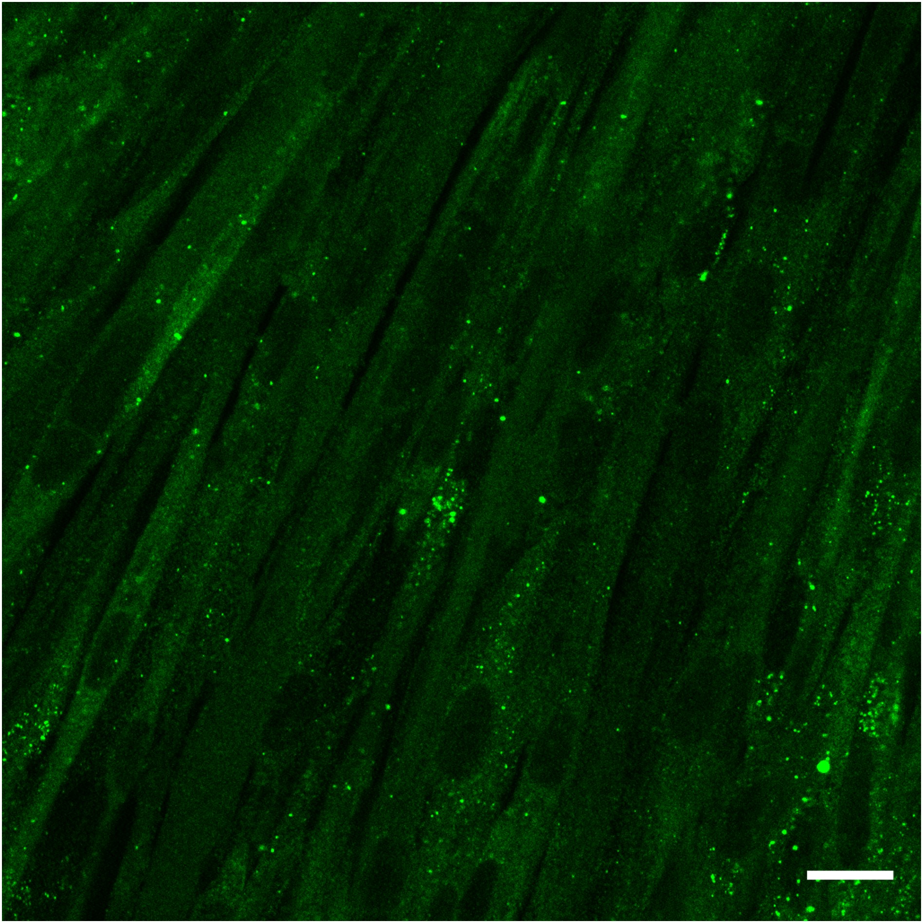 Immunostaining of p62 in DMD myotubes derived from patient cells under basal conditions, without autophagy activation.
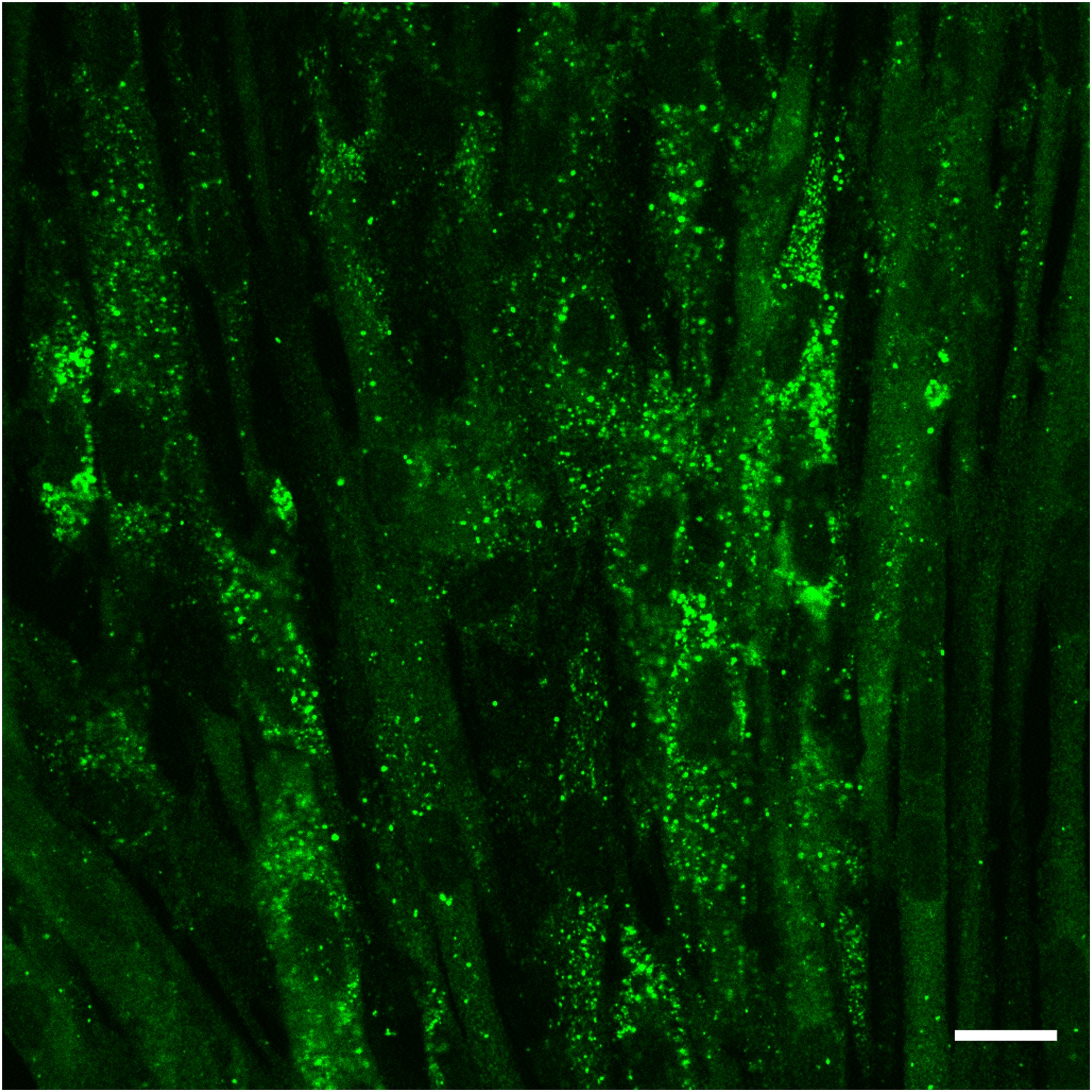 Immunostaining of p62 in the same model following induction of autophagy.
Selecting robust readouts
Work performed within DREAMS highlights the importance of selecting readouts that are both technically reliable and biologically meaningful.
Rather than relying on a single marker, multiple candidate readouts are evaluated in parallel. These include indicators of autophagy and lysosomal function, as well as additional phenotypic features identified across the diseases under study.
In some cases, alternative readouts are considered to complement autophagy measurements, particularly when they are already well-established in specific disease contexts. This approach helps ensure that assay development can proceed even when initial strategies require adaptation.
A key objective is to identify readouts that are compatible with screening approaches while retaining a meaningful link to the underlying biology. In practice, this requires balancing experimental scalability with biological relevance. High-throughput assays often rely on simplified or indirect measurements, which can facilitate comparisons across conditions but may only partially reflect the complexity of the autophagy pathway.
In this context, selecting appropriate readouts involves not only technical considerations, such as robustness, reproducibility, and sensitivity, but also a clear understanding of what aspect of the process is being captured. Readouts that are easily quantifiable may not always provide sufficient insight into autophagic flux or pathway dynamics, highlighting the need for complementary approaches.
Working with iPSC-derived muscle cells
A central component of the DREAMS approach is the use of skeletal muscle cells derived from induced pluripotent stem cells. These models provide a controlled system in which disease-related processes can be studied in a consistent manner.
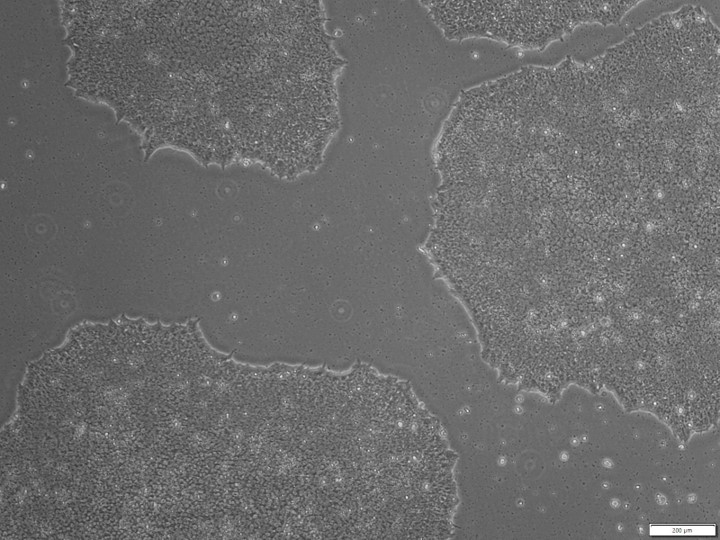
Image of DNM2 iPSC cells organized in clusters (islets). Scale bar: 200 µM.
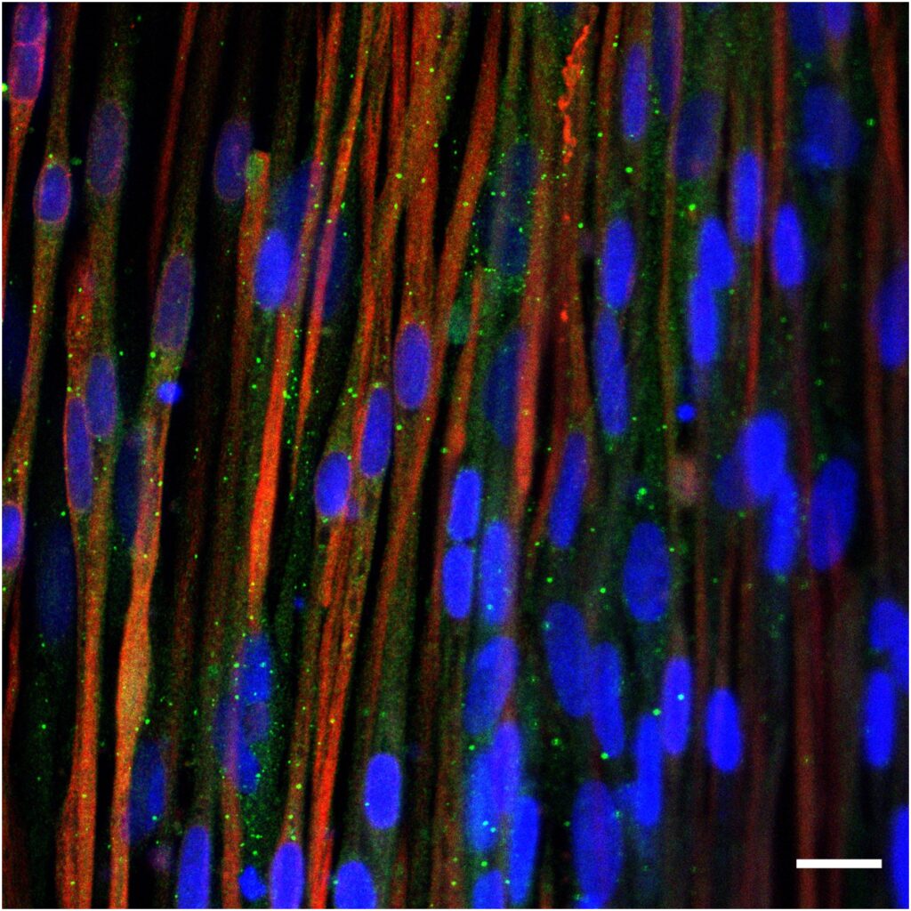
WT cells
DMD cells
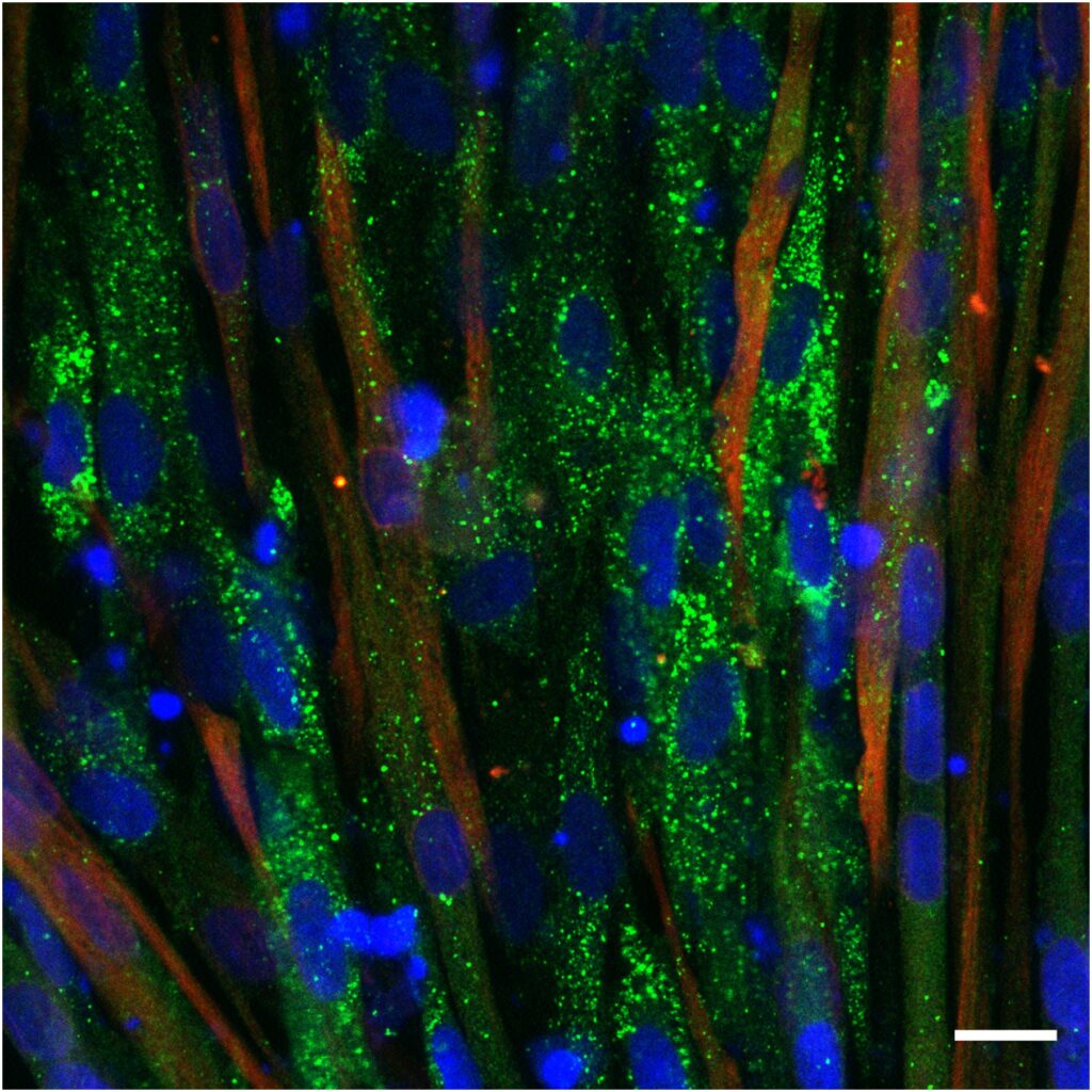
Image of myotubes derived from patient and healthy donor iPSC cells after autophagy activation, stained for an autophagy marker (green), Desmin to visualize myotubes (red), and nuclei (blue). Scale bar: 200 µM. Magnification: 20X.
iPSC-derived myotubes following pharmacological activation of autophagy, stained for an autophagy marker (green), Desmin to visualize myotubes (red), and nuclei (blue). Scale bar: 200 µM. Magnification: 20X.
At the same time, these models introduce additional variables that need to be carefully controlled during assay development. Distinct cellular states, such as myoblasts and myotubes, exhibit different basal autophagy levels and differential responses to modulation. Moreover, differentiation stage, culture duration, and fusion efficiency must be tightly controlled to ensure reproducible measurements.
Accordingly, assay design requires not only the selection of appropriate readouts, but also the careful choice of cellular model and differentiation stage. Experimental conditions must be optimised to achieve a balance between physiological relevance and signal robustness.
From assay development to screening
Once candidate readouts have been identified, they need to be adapted to formats compatible with high-throughput screening.
In DREAMS, this involves scaling assays to multi‑well formats and validating their performance using appropriate controls and statistical parameters such as Z’‑factor, signal‑to‑background ratio, and coefficient of variation. The use of known modulators of autophagy allows the identification of reference conditions and supports the selection of suitable readouts.
Pilot experiments indicate that compounds can have variable effects depending on the selected measurement. This reinforces the need to combine multiple readouts and to interpret results within the context of the assay design.
Integrating assays into a broader research pipeline
The assays developed in DREAMS are part of a broader framework that combines experimental and computational approaches.
Data generated from cellular models and screening experiments, including imaging‑based readouts, transcriptomic profiles, and proteomic analyses, contribute to the identification of shared pathways across different neuromuscular disorders. These datasets are then used to support further analysis, including approaches based on artificial intelligence.
In this context, the consistency and quality of the experimental data are essential. Well-characterised assays provide a foundation for generating datasets that can be compared across models and reused in different stages of the project.
Towards a structured analysis of autophagy
Rather than treating autophagy as a single measurable parameter, the approach followed in DREAMS considers it as a process that requires multi-dimensional analysis.
This involves combining different types of readouts, adapting assays to relevant cellular models, and validating measurements under controlled conditions. By doing so, the project aims to improve the consistency of experimental observations and to support a more structured understanding of disease-related mechanisms.
This work contributes to ongoing efforts to study neuromuscular disorders in a comparative and data-driven way, where the interpretation of biological processes depends on the reliability of how they are measured.
Meet the expert
Juliette Lemoine
PhD, Research Associate at I-Stem
References:
